

Scientists have long studied biological systems — first exploring, then exploiting, their natural elegance. In immunology, biological explorers were captivated by the workings of antibodies, proteins that seek and neutralize microbial threats.
Antibodies turned out to be fast-changing and nimble components of a natural defense system, and this versatility presented an opportunity to create a seemingly boundless array of medicines and clever research tools.
But how did this journey from investigation to innovation happen?
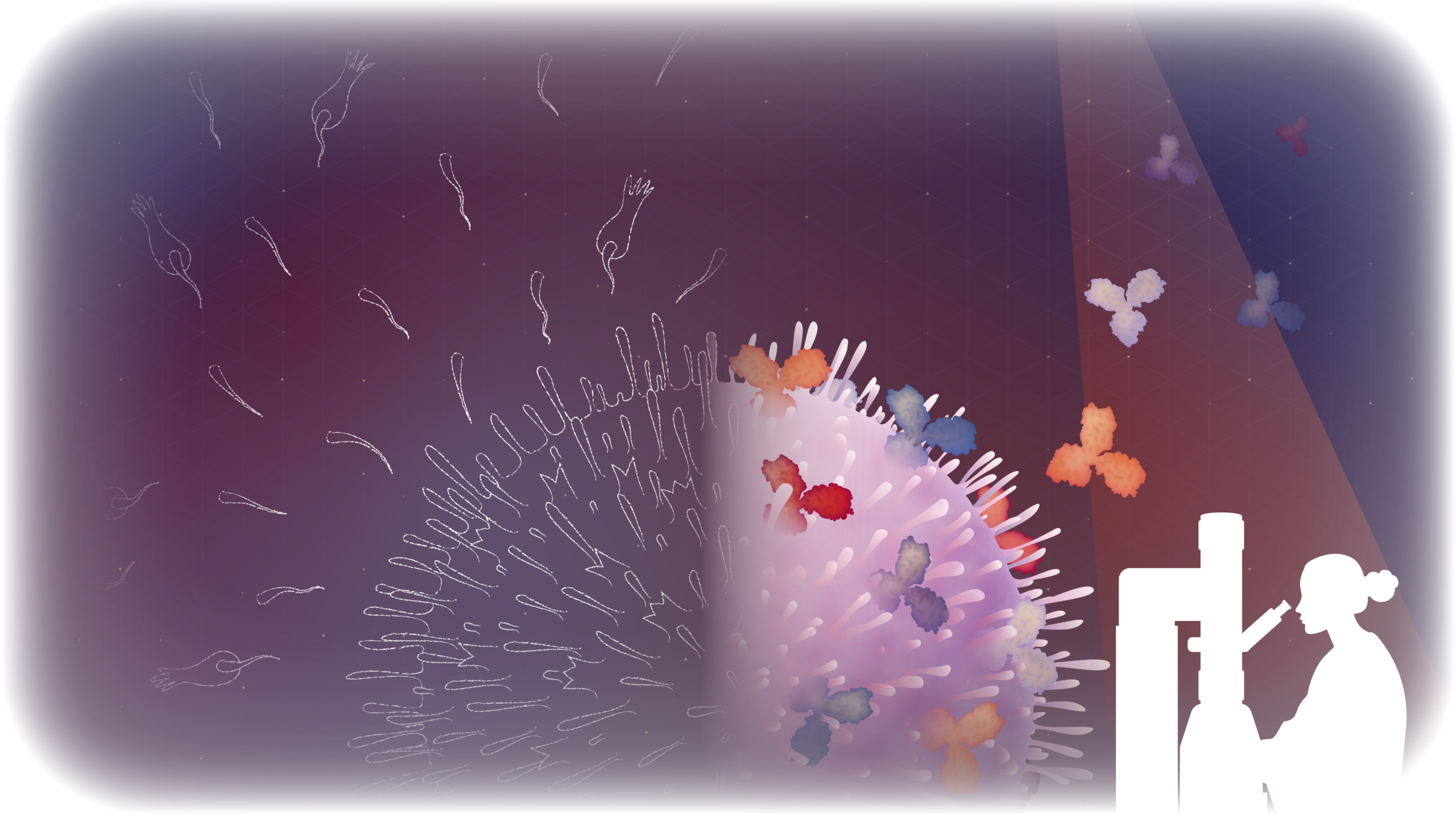
The birth of immunology
Our story begins in the 18th century, when Edward Jenner poked and pinched fluid out of a cowpox pustule, successfully vaccinating a young boy. The feat marked an important milestone in the advent of immunology, which took off in 1890 when Emil von Behring and Shibasaburo Kitasato discovered blood-borne “antitoxins,” or “stable properties” that disarmed diphtheria and tetanus toxins.
For this work, von Behring won the Nobel Prize in Physiology or Medicine in 1901 (Kitasato’s work was nominated but went unrecognized). The Prize committee noted “serum therapy” opened a limitless new field of medical science and “placed in the hands of the physician a victorious weapon against illness and death.”
It was German bacteriologist Paul Ehrlich, though, who coined the term “antibodies,” giving substance to the elixir in the serum that conferred immunity. He suggested that blood cells produced and shed the so-called antibodies when confronted with harmful toxins.
Though his hypotheses weren’t yet entirely correct, his work was honored by the Nobel Committee in 1908 and did inch us closer to our current understanding, while adding a new dictionary entry.
Nature’s defense mechanism
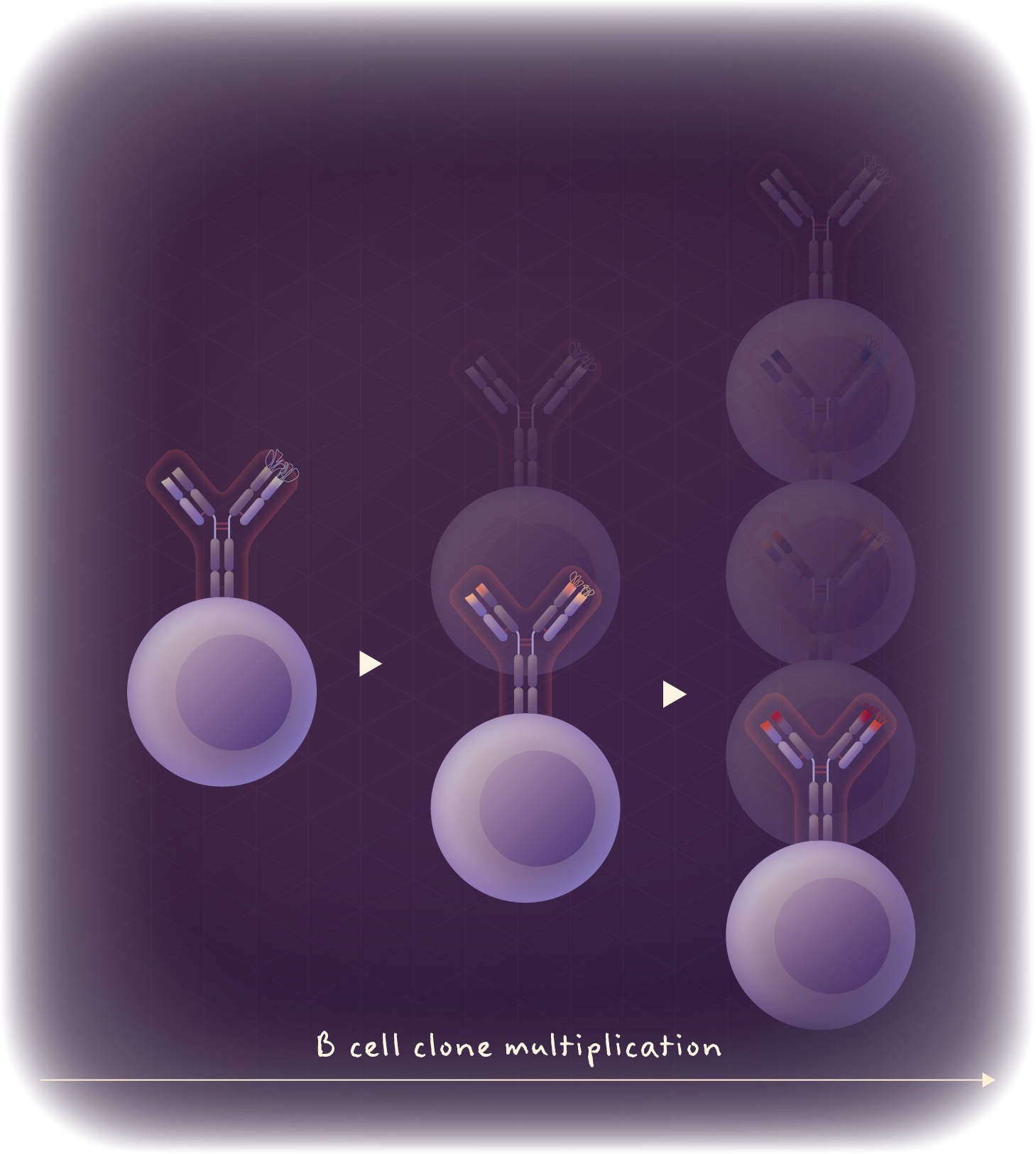
Today, we recognize that immune cells called B lymphocytes (B cells) are responsible for producing a vast repertoire of antibodies ready to recognize whatever target, or antigen, may come along.
Hover to change view.
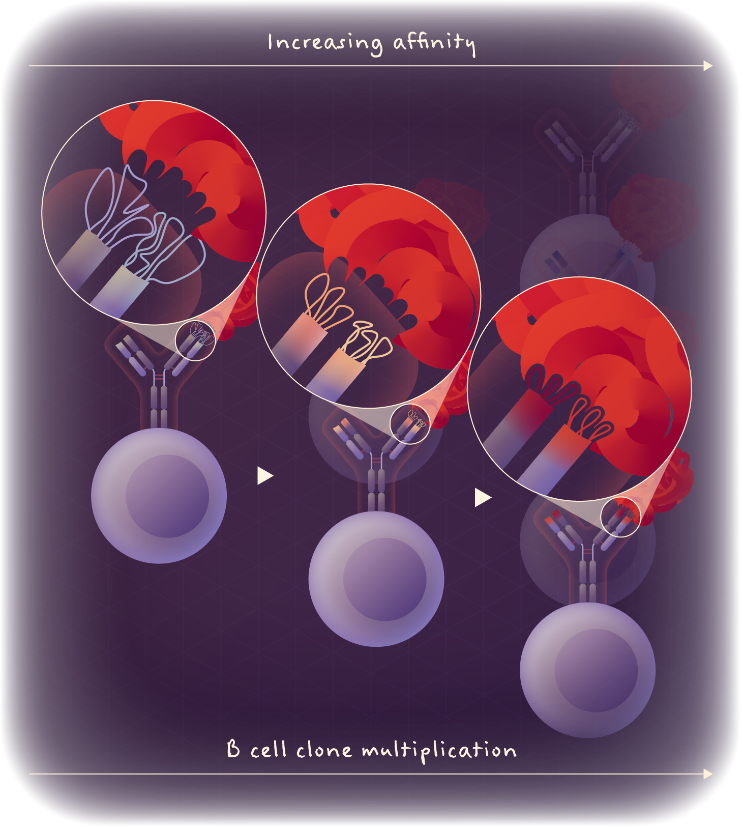
Each B cell typically expresses just one type of antibody, called a B cell receptor (BCR), and together the fleet produces billions of BCRs (the exact quantity is unknown but exceeds 1010). Each has a different antigen-binding site, or paratope, and, therefore, exhibits different binding preferences.
When a BCR binds to an antigen, the B cell carrying that antibody proliferates, generating many cells with many copies of the same antibody. The cells’ antibody DNA will rapidly mutate in a process called affinity maturation, producing slightly altered antibodies which more tightly bind their antigens.
There’s certainly more to it than that. But through more than a hundred years of work, this is our basic understanding of how the immune system selects and optimizes antibodies that can bind to antigens that pose a threat.
Polyclonal antibody production
Innovation generally follows understanding, and soon immunologists were thinking up ways to co-opt this natural immunity.
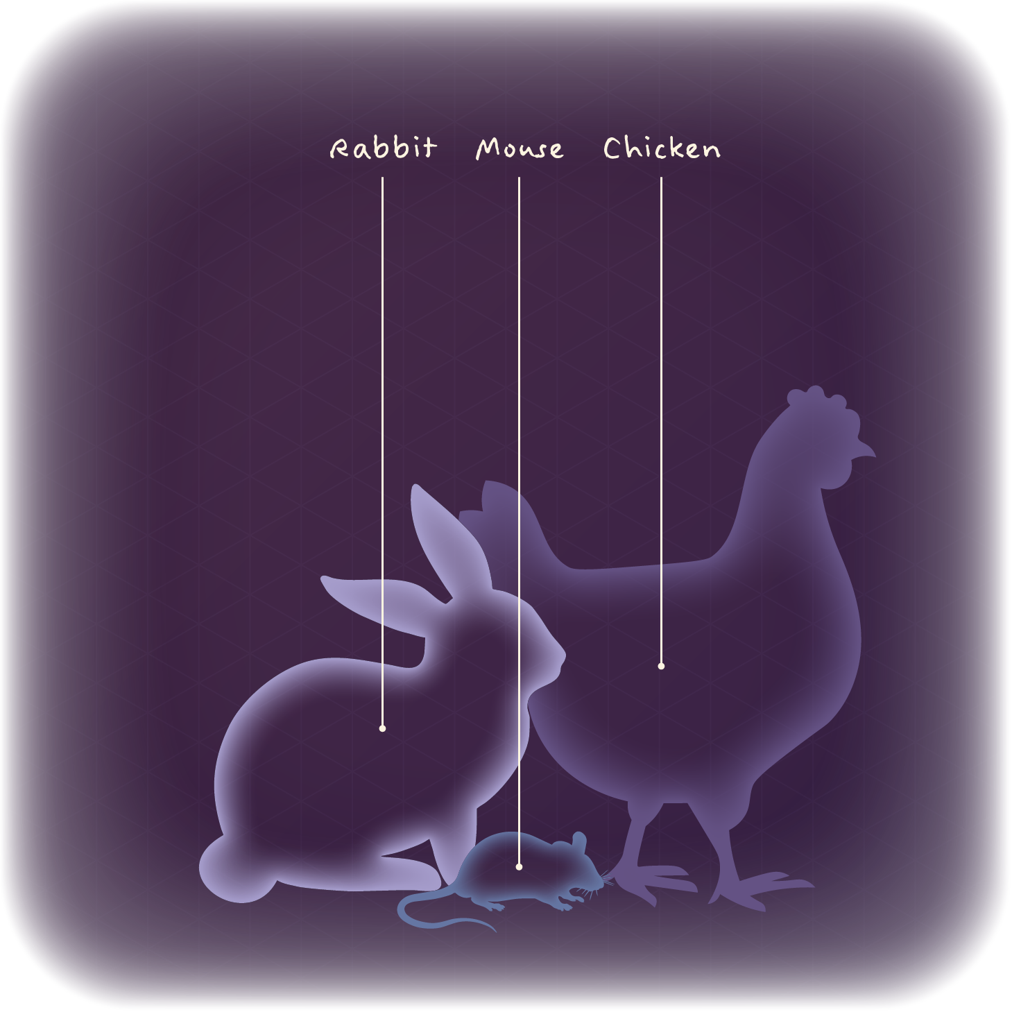
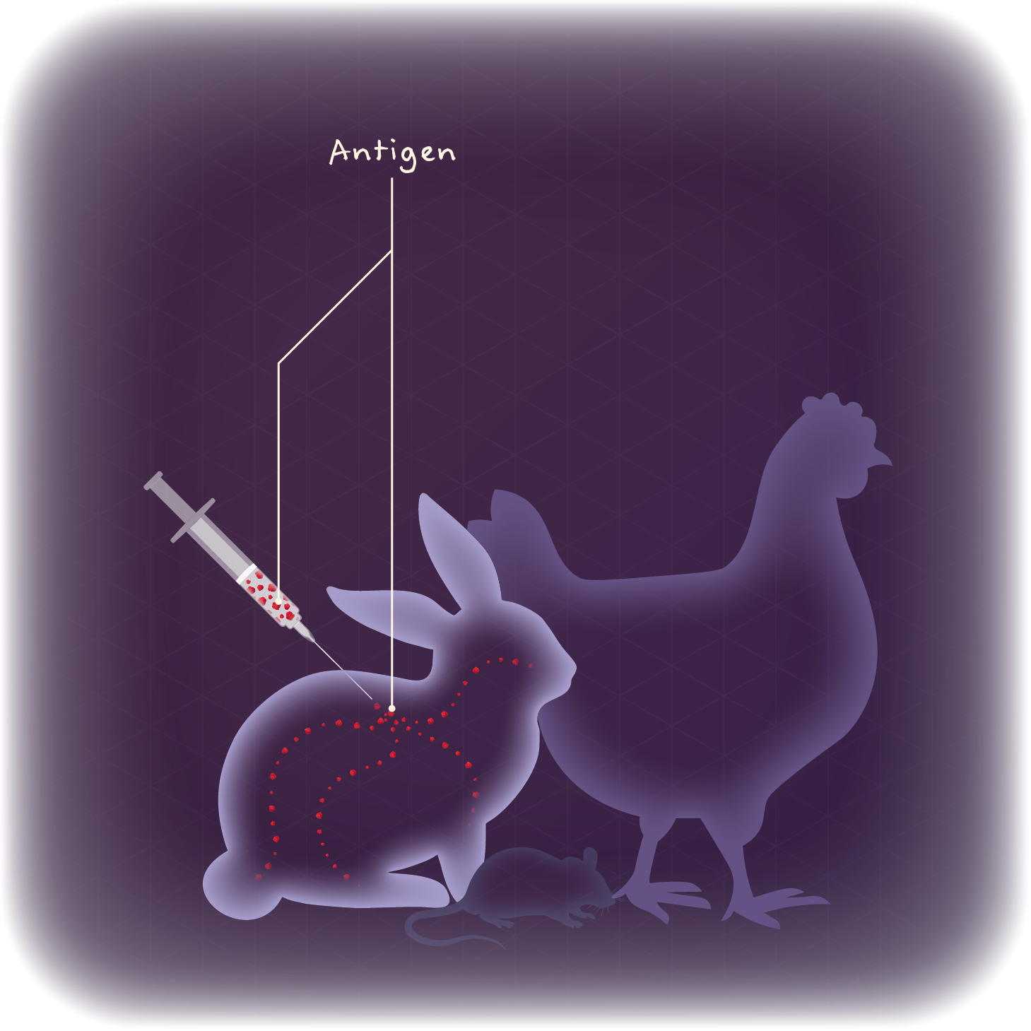
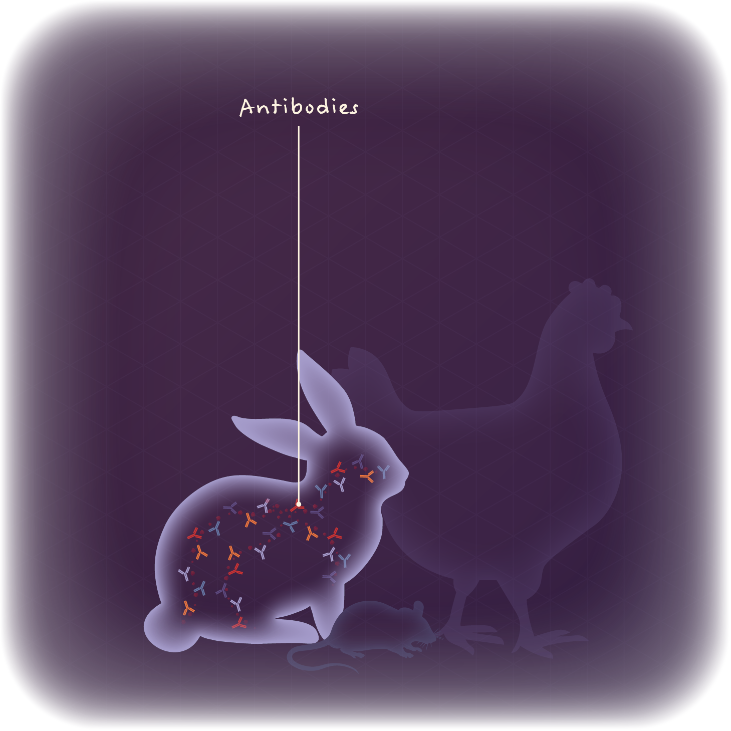
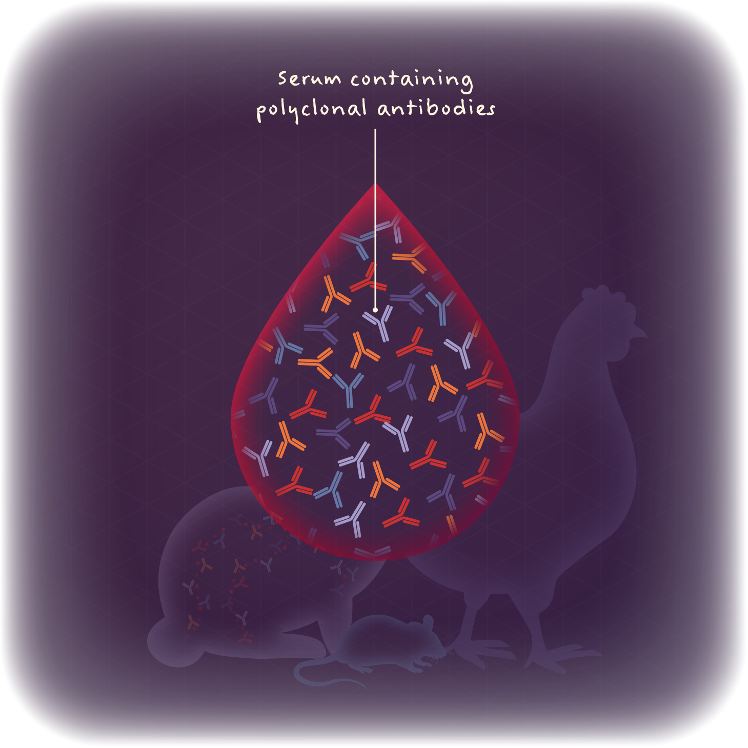
The lowest-hanging fruit came in the form of animal inoculation. Modern-day scientists inject an animal — often a mouse, rat, rabbit, or llama — with their protein of interest, then collect and purify the resulting antibodies from the animal’s blood serum. The method is similar to that practiced by Ehrlich and others, now standardized for industrial-scale production.
Antibodies produced in this way are called polyclonal antibodies, because they bind to a wide array of epitopes, or binding sites, on their targets — hence “poly.”
Think of it like a volunteer fire brigade, with many untrained, non-specialists working together. The antibodies are divergent, not directed against a specific epitope, but they get the job done nonetheless.
Polyclonals do perform well in some applications, particularly experiments where the precise makeup or conformation of an antigen is unknown or changing. But in many cases, the antibodies’ lack of specificity and reproducibility are problems. Even if scientists introduce an antigen into the same animal, its immune system will respond with a different set of antibodies. This means the exact cocktail cannot be replicated, and once the material runs out, it’s gone.
Hover to pause animation.
Monoclonal antibody technology
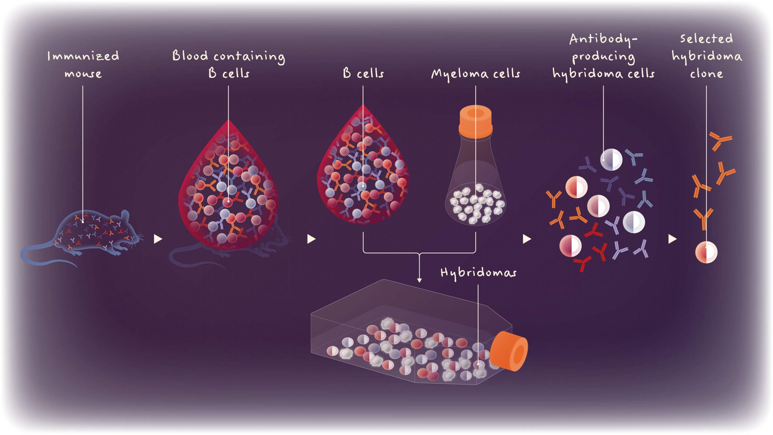
In 1975, researchers Georges J. F. Köhler and César Milstein broke ground with a stunning new way to make specific antibodies from a single B cell clone, so-called monoclonal antibodies. Their trick was to take antibody-producing B cells from an immunized animal and fuse them with immortal cancer cells, yielding “hybridoma” cells that could produce the encoded antibodies in large quantities.
From a pool of many hybridomas, scientists could select the clones that produced the best-binding antibodies. And because the antibodies came from identical clones, the collection targeted a single epitope on the antigen — hence “mono.” Once researchers devised how to maintain these hybridomas in culture, it became possible to churn out their monoclonal antibodies indefinitely. Or so it seemed.
As hybridoma technology became more widespread, researchers experienced a few notable problems. As the cells divided, for example, their DNA sequences would mutate, rendering monoclonal antibodies unreliable over longer timespans. At the same time, different lots of the same antibody crop could deviate, with a noticeable impact on experimental results.
The resulting inconsistencies have cast doubt on many credible scientists’ studies, stained the reputation of otherwise impeccable work, and contributed to a “reproducibility crisis” in biomedical research.
The recombinant revolution
In the 1970s and ’80s, advances in genetic engineering began to drive further innovation. One enormous step came when two researchers in California — Stanley Cohen and Herbert Boyer — developed recombinant DNA technology, which allowed scientists to manipulate DNA fragments from different genes and species. This technology opened new doors in biotechnology and pharmaceutical development, and revolutionized antibody production, too.
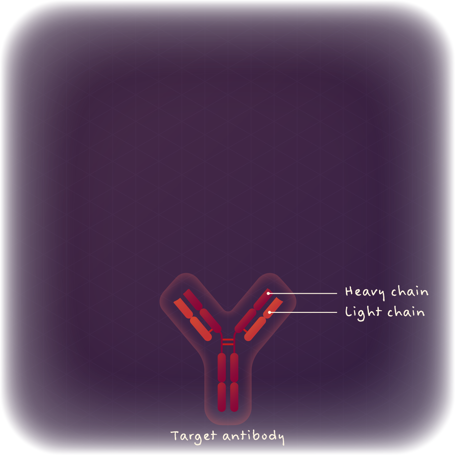
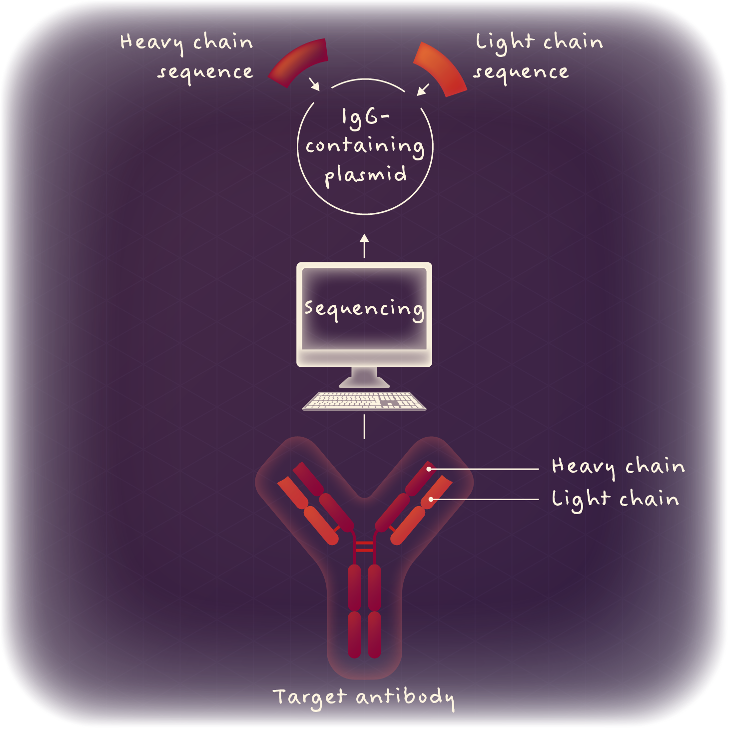
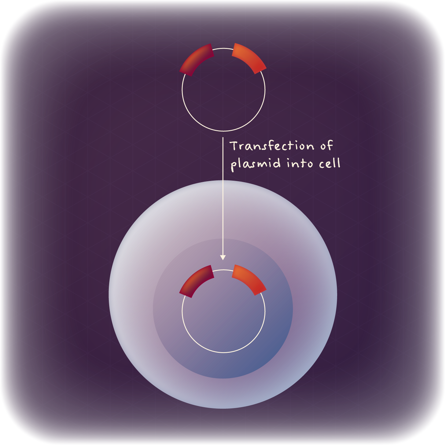
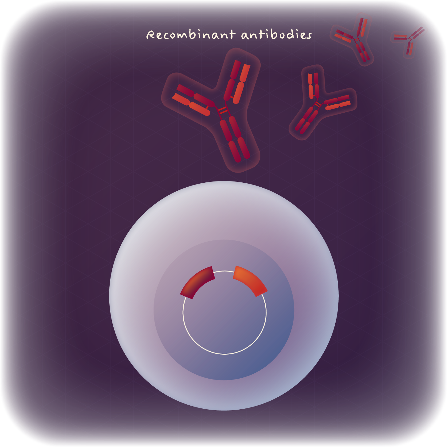
Using this tool, scientists could extract and sequence the DNA encoding a prized monoclonal antibody, paste the sequence into a vector, and then express that antibody fragment in a more controlled, high-expressing cell line — like human embryonic kidney (HEK) cells or Chinese hamster ovary (CHO) cells — before purification.
Hover to pause animation.
Antibodies made in this way were called recombinant antibodies. Due to their grounded DNA sequences, they were highly consistent from batch to batch, unlike traditional hybridomas. Plus, they were easily customizable. For example, antibody domains from two different species with enviable binding traits could be combined to form a chimeric antibody that demonstrated optimal, and previously impossible, binding.
Initially, recombinant antibodies were hybridoma-derived, relying on the front-end production of monoclonal antibodies through animal immunization. Though this is still a common starting point for recombinants (including a cluster of integrin antibodies at IPI), scientists in the 1990s began to generate fully synthetic recombinant antibodies — entirely in vitro.
Still getting to know antibodies? Start at the beginning of our Explainer series.
Writers: Caitlin Faulds, caitlin.faulds@proteininnovation.org
Megan Talkington contributed to this story.
Illustrator: Anne-Lise Paris, www.in-graphidi.com
About IPI
The Institute for Protein Innovation is pioneering a new approach to scientific discovery and collaboration. As a nonprofit research institute, we provide the biomedical research community with synthetic antibodies and deep protein expertise, empowering scientists to explore fundamental biological processes and pinpoint new targets for therapeutic development. Our mission is to advance protein science to accelerate research and improve human health. For more information, visit proteininnovation.org or follow us on social media, @ipiproteins.